


Osteoarthritis (gonarthrosis) is a pathological alteration of the knee joint that has a chronic course and can progress over time. The disease extends to all components of the knee joint: cartilage, subchondral bone, menisci, synovial membranes, ligaments, capsules and periarticular muscles.
The knee joint, which connects the femur and tibia, is subjected to heavy loads throughout life and is regularly injured. Sometimes minor damage immediately goes unnoticed, but makes itself felt in the second half of life. In this regard, joint damage is often detected in the elderly. However, even young people suffer from knee joint disorders resulting from sports or an active lifestyle.
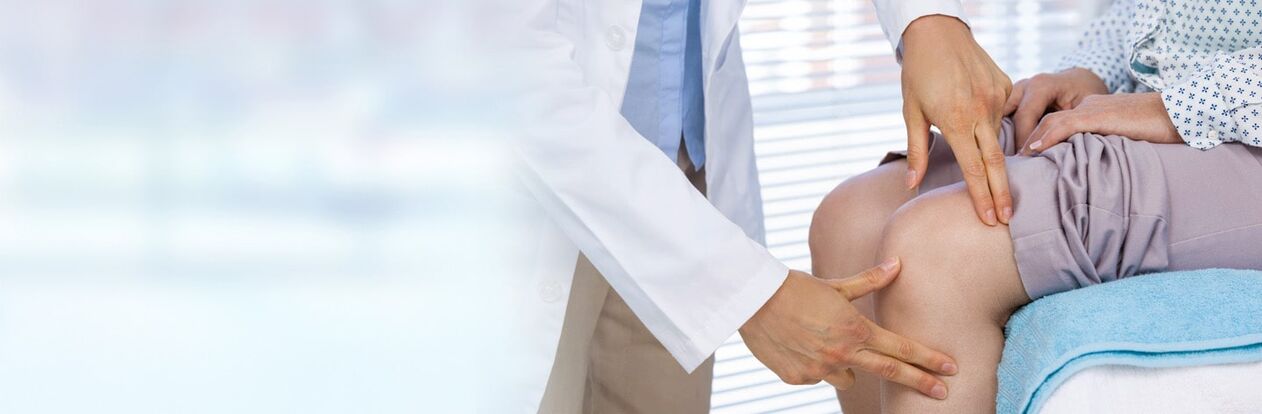
To maintain maximum mobility and a high standard of living, you should consult a doctor at the first problem with the knee joint. Experienced orthopedic traumatologists will diagnose your condition and prescribe the necessary treatment.
Types of arthrosis of the knee joint
Inside the joint, the bones are covered with cartilaginous tissue, which provides shock absorption, smoothness, and also prevents the bones from rubbing against each other. The cartilaginous tissue receives nourishment thanks to the synovial fluid located inside the joint and the blood that flows through the vessels.
Cartilage has a spongy structure, so when it is at rest it absorbs fluid and when it is loaded it moves it. At the same time, during movements, the cartilage constantly receives microtraumas, and during rest it is restored.
If the result of a mechanical injury exceeds the ability of the joint to restore, there is not enough nourishment for the cartilage and, as a result, regeneration does not occur. The damage accumulates and changes the structure of the cartilage tissue. This is how arthrosis of the knee joint begins.
Depending on the reasons that provoked it, arthrosis of the knee joint is usually divided into two types: primary and secondary.
Primary gonarthrosis
Degenerative changes that occur in the joint are associated with age. Among the reasons that cause it are the following:
- natural degeneration or degradation due to the slowing down of metabolic processes in the body;
- excess body weight;
- sedentary lifestyle;
- malnutrition;
- genetic predisposition.
As a rule, primary gonarthrosis affects both knees at the same time and is called bilateral.
Secondary gonarthrosis
Secondary arthrosis of the knee joint can occur at any age, as it can be caused by:
- various injuries - contusions, fractures, dislocations, ruptures and sprains of ligaments or menisci;
- joint diseases: rheumatoid arthritis, osteochondritis dissecans, gout, goniitis, etc. ;
- regional vascular disorders;
- overload of the knee joints during sports or due to the specifics of work;
- endocrine diseases;
- O-shaped and X-shaped leg curvature.
Secondary arthrosis of the knee joint most often appears only on one leg and is called unilateral.
In rare cases, idiopathic gonarthrosis is identified, a disease that occurs for no apparent reason.
Stages and symptoms of arthrosis of the knee joint
Regardless of how arthrosis of the knee joint appeared, experts distinguish three stages of its development, which are determined during an x-ray examination. Each phase is accompanied by characteristic symptoms:
Phase 1– mild pain that occurs after prolonged exercise, when climbing stairs, after intense physical exercise and disappears after rest. There are no restrictions on movement, but sometimes mild swelling of the joint may occur. This condition, if nothing is done, can last for years: at this time the cartilage is just starting to lose its smoothness due to impaired blood supply. An x-ray will show slight narrowing of the joint space and hardening of the bones.
- Phase 2– the pain becomes intense and lasts a long time even with light effort. A creaking sound is heard during flexion and extension of the joint. It becomes impossible to bend the leg completely due to severe pain. There is slight deformation, muscle atrophy and limited movement. The pain can be relieved with painkillers or disappear on its own after rest.
At this stage the cartilaginous layer is already thinning considerably, until it disappears in some places. Synovial fluid becomes thicker and more viscous, which compromises its nutritional and lubricating properties. Osteophytes appear: bone growths.
- Phase 3– the pain increases and worries constantly, even at night. The deformity of the joint becomes noticeable, the gait changes and the lower part of the limb bends. The range of motion of the knee joint is reduced: the leg cannot be bent or straightened completely. When walking, you need to use support in the form of a cane or crutch. Painkillers no longer help.
The cartilage is almost completely erased, the bones are compacted, the joint space is noticeably narrowed or absent. The presence of numerous osteophytes is noted.
A common symptom of arthrosis of the knee joint can be identified: pain of varying intensity, localized along the anterior-internal surface of the joint.
Diagnostics
If you observe symptoms similar to the development of gonarthrosis, you should consult a doctor. At the first visit, the doctor will take your medical history, check the biomechanical capabilities of the joint and prescribe the necessary tests. Be sure to inform him about injuries and illnesses suffered, lifestyle, diet, medications taken and job characteristics.
The most informative and simple way to confirm or refute a diagnosis is an x-ray of the knee joint: it allows you to conduct a differential diagnosis, determine the degree of development of arthrosis and monitor the treatment process.
However, radiological signs appear much later than morphological changes. Therefore, in the early stages, gonarthrosis is difficult to detect even with an x-ray. In such situations, the doctor may prescribe arthroscopy - a highly accurate method of diagnosing changes in the joints using special endoscopic equipment.
Additional research methods are ultrasound and magnetic resonance imaging: they are prescribed when the x-ray is not sufficiently informative.
Treatment of arthrosis of the knee joint
After the diagnosis, the doctor selects the optimal treatment, depending on the stage of the disease and individual characteristics. This solves three problems:
- pain relief;
- stop the progression of the pathology;
- restoration of joint function.
The specialist selects a comprehensive solution, which can be adapted during the treatment process.
In modern medicine there are many ways to treat joint diseases. They can all be divided into three types: conservative, minimally invasive, surgical.
Conservative method of treatment of gonarthrosis
Usually used in stages 1-2 of arthrosis of the knee joint. Treatment begins with reducing the load on the joint: the patient must avoid excessive vertical load on the joint: jumping, running, etc. If necessary, it is recommended to lose excess weight. The doctor will recommend a diet and select a series of gentle exercises that will reduce axial impacts and improve the nutrition of the cartilage tissue.
To improve blood circulation in the joint area, increase freedom of movement, and also improve the effect of drugs, physiotherapy is prescribed:
- shock wave therapy – short-term impact on bone and connective tissue with acoustic pulses of significant amplitude at low frequency;
- electrotherapy – exposure of the affected area to electric current, magnetic or electromagnetic fields;
- laser therapy: exposure to optical radiation generated by a laser;
- phonophoresis: exposure of the affected area with ultrasound and medicines applied to the skin;
- electrophoresis: exposure of the affected area to electricity.
Massages, compresses, the use of orthoses and kinesiotaping have also proven effective in treating osteoarthritis.
In addition, well-chosen drug therapy helps to relieve pain, stop inflammation and slow down the process of destruction of cartilage tissue. For this purpose, anti-inflammatory, hormonal, antispasmodic and chondroprotective drugs are prescribed. They can be in tablet, injectable or topical form, depending on the situation.
Minimally invasive method for the treatment of gonarthrosis
If the above procedures have no effect, your doctor may prescribe intra-articular injections:
- hyaluronic acid – replaces synovial fluid to improve friction, reduce pain and improve knee joint function. The average duration of action of the drug is 3-6 months;
- own plasma enriched with platelets - for nutrition and restoration of cartilaginous tissue;
- corticosteroids – to reduce inflammation.
Surgical method for the treatment of gonarthrosis
If conservative treatment proved ineffective or you first turned to a specialist with the third stage of arthrosis of the knee joint, the doctor may resort to surgical intervention:
- arthrodesis: artificial immobilization of the affected joint in a physiological position to eliminate pain;
- arthroscopic debridement: joint hygiene using an arthroscope;
- corrective osteotomy – elimination of bone deformities by artificial fracture;
- endoprosthesis: replacement of a worn joint with an implant created artificially with biocompatible materials.
The type of intervention is chosen by the doctor based on the characteristics of the arthrosis of the knee joint. But endoprosthesis is considered the gold standard, as it allows you to completely return to your normal lifestyle. At the same time, a good plant does not require replacement for 15-30 years. To fully recover after surgery, it is necessary to undergo a rehabilitation process that lasts 3-4 months.
Complications
Gonarthrosis develops quite slowly, but it can be detected timely and the necessary treatment can begin. Ignoring the disease and its symptoms can lead to serious consequences:
- constant pain that is not relieved by medications;
- complete immobility of the diseased joint;
- inability to lean on the injured limb;
- severe joint deformation and curvature of the legs;
- damage to other parts of the musculoskeletal system;
- shortening of the leg.
In particularly difficult situations and in the absence of timely treatment, arthrosis can lead to disability and deterioration of motor activity, up to immobility.
It is important to remember that it is impossible to completely cure arthrosis. But it is quite possible to stop the progression of the disease and improve the quality of life.
Prevention
There is no preventive treatment for gonarthrosis. But people at risk are advised to adhere to certain rules:
- make sure that your weight does not exceed the age norm;
- do not practice sports that place great stress on the knee joint;
- if possible, completely cure infectious diseases without causing complications;
- don't get too cold or too tired;
- avoid injuries and overloading of the joint;
- avoid stressful situations;
- do not forget about rest;
- engage in physical therapy;
- wear orthopedic shoes.
At-risk groups include older adults, athletes and dancers. You can also add here those who lead a sedentary lifestyle, stand a lot at work or lift weights and are overweight.
Any change in the axis of the lower limb or in the normal biomechanics of the joint, dysplasia, decreased volume and strength of the leg muscles, or trauma can also lead to osteoarthritis.
Get checked regularly and take preventative measures.
Question Answer
- What is the difference between knee arthritis and knee osteoarthritis?
Arthritis is the collective name for inflammation of the joint and arthrosis is a degenerative-dystrophic process.
- Which doctor treats osteoarthritis?
Traumatologist-orthopedist or rheumatologist.
- Is it possible to play sports with arthrosis of the knee joint?
Avoid prolonged and heavy loads on the joint, as well as axial shocks. But you should not completely exclude sport from your life: when you move, your joints are better "nourished" and restored. It is important to observe the measure and adhere to the recommendations of the doctor, who will select the type and mode of exercise.